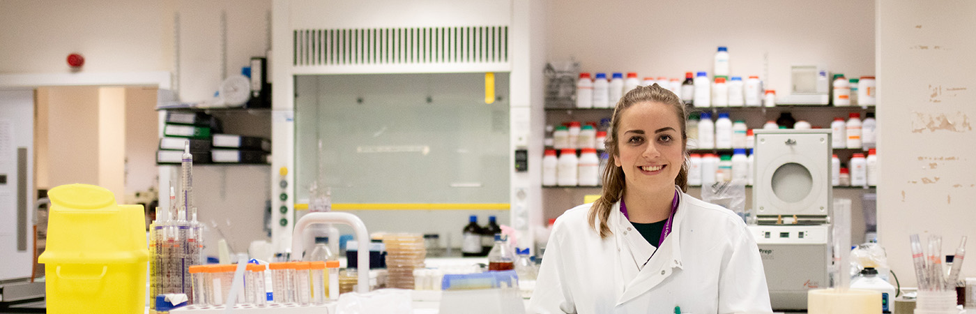
Donor-funded PhD student Victoria is producing a novel way to combat Keratitis, a sight-threatening eye disease.
PhD student Victoria has modelled an eye in a petri dish to conduct more accurate research into an eye disease Share on X Thanks to donations, Victoria is able to carry out groundbreaking research into Keratitis Share on X Find out how donor funding is enabling research that has a real, positive impact on the global population Share on XThe use of the petri dish has been commonplace in science for over 100 years. Many of us will have used one in school to carry out basic experiments like assessing the growth of mould. What we may not have envisaged however is that something much more complex could be grown in a petri dish. A human eye, for example.
That is the aim for PhD student Victoria Rimmer. She is creating a model of the cornea, the surface of the eye, in a petri dish in order to carry out more accurate research on a disease called keratitis.
Pseudomonas aeruginosa keratitis refers to sight-threatening inflammation of the cornea that is often caused by an infection. Infection with the bacterium Pseudomonas aeruginosa is common in contact lens wearers and in rural areas of the developing world where access to the right drug treatment is limited.
“If not treated properly, keratitis can lead to blindness. There are more and more people now wearing contact lenses and they can be quite unhygienic with them; they don’t clean them properly, they shower in them, sleep in them, and the surface of the lens can cause bacteria to build up near the surface of your eye and that can increase your risk of getting a corneal infection.”
A remodelling of the ocular surface
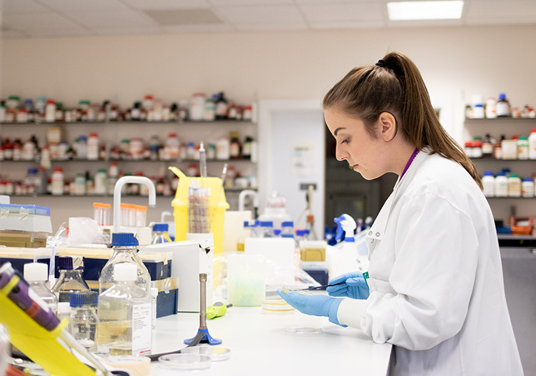
Victoria, a recipient of a donor-funded Research Impact Scholarship, is putting together an in vitro remodelling of the ocular surface using donated human corneas and tears. The process involves creating primary epithelial cell cultures from the corneas and also developing a real tear solution using tear proteins.
“We want to see how those tears and that model system impact bacteria, so we have some clinical Pseudomonas aeruginosa strains from patients who have eye infections and we’re going to challenge our model with those bacteria.
“We’re then hoping to understand how the presence of all those antimicrobials and the cells affect the behaviour of the bacteria; does it make them more or less likely to be able to cause an infection?”
Victoria has made a model of the cornea predominantly made of collagen and, on top of that, a layer of epithelial cells that forms a protective layer over the cornea that bacteria usually can’t penetrate.
“Normally what would then happen is you’d scratch your eye, for example, or you’d wear a contact lens that somehow changes that epithelial layer and allows the bacteria to get into your eye. The next step is to develop the tears so that we can put the tears on that model and see if they affect how the bacteria behave.”
A viable alternative to animal testing
Research on keratitis is not necessarily novel in itself, but the method and accuracy with which Victoria plans to conduct the research is completely unique.
“There’s research being carried out on keratitis but it is often done using mice or other animals. There is no complex model being created in the lab with the right tears and cells.”
Considerable efforts have been made in recent years to reduce the need for animal testing. However, the reality is that we are still reliant on animals for certain tests.
“We hope that the model will provide a reproducible alternative to animal models in the study of keratitis, and could be used as a pharmaceutical and therapeutic tool in the future to benefit patients.”
Donor Impact
Thanks to many donors to the University, Victoria is well on the way to successfully creating an innovative and ground-breaking model that will enable more accurate research on a debilitating disease.
“For me, what’s really interesting is that if we can see the bacteria change in the way they produce proteins and other molecules, those could potentially be targets for drugs to treat infection.
“I think that’s quite important because antibiotic resistance is becoming a big problem so, if we can find some novel targets that seem to be increasing in keratitis, that might be something that we could use the model for as well and understand more about how this disease progresses in people.”
At The University of Manchester, we pride ourselves on carrying out research that will have a real impact on the world and will benefit future generations as well as the current one. Thanks to donations from many generous donors, we are able to provide Research Impact Scholarships to PhD students who aim to address global issues and change the world for the better with their research. Victoria is just one of a number of PhD students who are beneficiaries of the donor-funded Research Impact Scholarship Programme.
“I definitely wouldn’t have been able to study for my PhD without this scholarship. Until I became a postgraduate student, I didn’t realise how expensive research was. Even small consumables cost so much money and you can go through a lot without realising how much it’s costing. Without these types of scholarships, I think a lot of PhD students wouldn’t be able to do what they want to do.
“It would be amazing to have an impact on so many people, from those who wear contact lenses to people in really tough circumstances. I think that’s the whole point in doing a PhD like this, and in studying science on the whole.”